Implanty ceramiczne z tlenku cyrkonu
Implanty – najlepsza metoda odbudowy zębów
Przy użyciu implantów można odbudować jeden lub więcej brakujących zębów lub zrekonstruować całkowity brak uzębienia. Implanty od dawna są najbardziej atrakcyjną formą protez zębowych. Zapewniają one funkcjonalność, wygodę i dobry wygląd tym samym poprawiając jakość życia i pewność siebie. Celem jest by ząb odbudowany na implancie nie różnił się w użytku i wyglądzie od naturalnych zębów. Poprzez obciążenie kości implantami zapobiega się również zanikowi kości w miejscu utraconych zębów. Zapobiegają też przemieszczaniu się sąsiednich lub przeciwstawnych zębów oraz umożliwiają utrzymanie prawidłowego zgryzu.
Pozostałymi opcjami odbudowy brakujących zębów są mosty i protezy ruchome. W niektórych przypadkach lukę po zębie można też zamknąć przesuwając sąsiedni zęby, co wymaga leczenia ortodontycznego.
Implanty ceramiczne
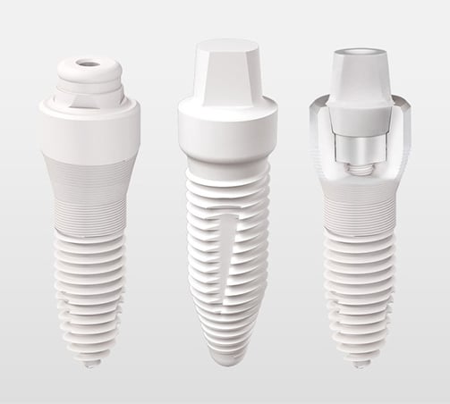
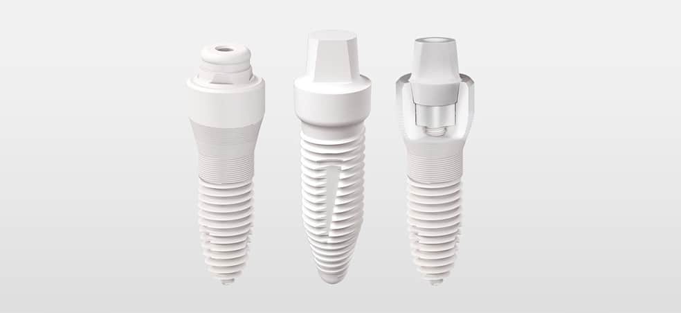
Niezależnie od tego czy odbudowywany jest jeden ząb, czy cały łuk, użyty materiał musi być stabilny i neutralny a do tego wytrzymały. Implanty ceramiczne wykonane są z tlenku cyrkonu (od dawna używanego też w endoprotezach stawu biodrowego), który całkowicie spełnia wymienione wymagania. Dzięki niezwykle stabilnej strukturze chemicznej, z ceramiki nie uwalniają się jony ani żadne inne związki chemiczne. Ceramika nie podlega korozji i nie wywołuje reakcji zapalnych. W naszej klinice używane są ceramiczne implanty cyrkonowe Szwajcarskiej firmy Swiss Dental Solutions (SDS), opracowane specjalnie z myślą o zdrowiu i estetyce. Implanty ceramiczne stanowią świetny wybór dla pacjentów pragnących trwale, minimalnie inwazyjnie i estetycznie odbudować swój uśmiech a także dla tych, którzy nie chcą wprowadzać metalu do organizmu lub mają nietolerancję tytanu.
Estetyczna odbudowa zębów
Piękne białe zęby i różowe dziąsła to wyraz zdrowia, energii i witalności. Wiemy jak znaczącym elementem wizerunku jest zadbany uśmiech i dlatego w naszej klinice wszystkie proponowane rozwiązania mają też na uwadze estetykę.
Cyrkonowe implanty ceramiczne są w całości białe – bardzo bliskie naturalnego koloru zębów. Dzięki temu, w przeciwieństwie do implantów tytanowych, nie ma ryzyka, że spod korony przebijać się będzie szary cień. Nie powstaną też szare krawędzie między dziąsłem a koroną zęba. Nawet jeżeli pokrywające dziąsło jest bardzo cienkie lub ulegnie recesji, implant zawsze pozostaje biały. Z tego powodu cyrkonowe implanty ceramiczne świetnie sprawdzają się w obszarze zębów przednich.
Zdrowiej bez metalu
Podczas gdy stosowanie metali w jamie ustnej może mieć negatywny wpływ na cały organizm, implanty ceramiczne z tlenku cyrkonu mają doskonałą kompatybilność, gdyż są w stu procentach bezmetalowe oraz immunologicznie obojętne. Ponadto, podczas implantacji używane są ceramiczne wiertła, żeby nie pozostawić w kości śladowych ilości metalu. Dzięki ich kompatybilności biologicznej, regeneracja dziąseł wokół implantu jest bardzo dobra. Ceramika pozwala też na wykonanie implantów ze specjalną strukturą powierzchni, która ogranicza gromadzenie bakterii i powstawanie płytki nazębnej, a tym samym ryzyko zapalenia dziąseł. Opatentowane struktury powierzchniowe SDS wraz z dostosowanymi do kształtu kości kształtami gwintu umożliwiają doskonałą integrację implantów, co oznacza, że zazwyczaj mogą one zostać poddane obciążeniu już po kilku tygodniach.
Badania i źródła do powyższego tekstu
Implanty ceramiczne
- Thoma DS, Lim H-C, Paeng K-W, Jung U-W, Hämmerle CHF, Jung RE. Tissue integration of zirconia and titanium implants with and without buccal dehiscence defects-A histologic and radiographic preclinical study. Clinical Oral Implants Research. 2019;30(7):660–9. doi:10.1111/clr.13451
- Beekmans DG. The pink and white aesthetics of a new zirconia implant. Nederlands Tijdschrift voor Tandheelkunde. 2018;125389–95. doi:10.5177/ntvt.2018.07/08.18134
- Hisbergues M, Vendeville S, Vendeville P. Zirconia: Established facts and perspectives for a biomaterial in dental implantology. J Biomed Mater Res Part B Appl Biomater. 2009;88(2):519–29. doi:10.1002/jbm.b.31147
- Fischer J, Benic G, Fischer Carolin. Zirkonoxidimplantate – wieso, weshalb, warum [Internet]. 2016. Available from: https://www.zmk-aktuell.de/fachgebiete/implantologie/story/zirkonoxidimplantate–wieso-weshalb-%20warum__4830.html
- Sivaraman K, Chopra A, Narayan AI, Balakrishnan D. Is zirconia a viable alternative to titanium for oral implant? A critical review. J Prosthodont Res. 2018;62(2):121–33. doi:10.1016/j.jpor.2017.07.003
- Manzano G, Herrero LR, Montero J. Comparison of clinical performance of zirconia implants and titanium implants in animal models: a systematic review. Int J Oral Maxillofac Implants. 2014;29(2):311–20. doi:10.11607/jomi.2817
- Özkurt Z, Kazazoğlu E. Zirconia dental implants: a literature review. J Oral Implantol. 2011;37(3):367–76. doi:10.1563/AAID-JOI-D-09-00079
- Payer M, Heschl A, Koller M, Arnetzl G, Lorenzoni M, Jakse N. All-ceramic restoration of zirconia two-piece implants–a randomized controlled clinical trial. Clinical Oral Implants Research. 2015;26(4):371–6. doi:10.1111/ clr.12342
- Möller B, Terheyden H, Açil Y, Purcz NM, Hertrampf K, Tabakov A, Behrens E, Wiltfang J. A comparison of biocompatibility and osseointegration of ceramic and titanium implants: an in vivo and in vitro study. Int J Oral Maxillofac Surg. 2012;41(5):638–45. doi:10.1016/j.ijom.2012.02.004
- Koch FP, Weng D, Krämer S, Biesterfeld S, Jahn-Eimer- macher A, Wagner W. Osseointegration of one-piece zirconia implants compared with a titanium implant of identical design: a histomorphometric study in the dog. Clinical Oral Implants Research. 2010;21(3):350–6. doi:10.1111/j.1600-0501.2009.01832.x
- Kohal RJ, Weng D, Bächle M, Strub JR. Loaded custom-made zirconia and titanium implants show similar osseointegration: an animal experiment. J Periodontol. 2004;75(9):1262–8. doi:10.1902/jop.2004.75.9.1262
- Roehling S, Schlegel KA, Woelfler H, Gahlert M. Zirconia compared to titanium dental implants in preclinical studies- A systematic review and meta-analysis. Clinical Oral Implants Research. 2019;30(5):365–95. doi:10.1111/clr.13425
- Bormann K-H, Gellrich N-C, Kniha H, Schild S, Weingart D, Gahlert M. A prospective clinical study to evaluate the performance of zirconium dioxide dental implants in single-tooth edentulous area: 3-year follow-up. BMC Oral Health. 2018;18(1):181. doi:10.1186/s12903-018-0636-x
- Hashim D, Cionca N, Courvoisier DS, Mombelli A. A systematic review of the clinical survival of zirconia implants. Clin Oral Investig. 2016;201403–17. doi:10.1007/s00784- 016-1853-9
- Roehling S, Schlegel KA, Woelfler H, Gahlert M. Performance and outcome of zirconia dental implants in clinical studies: A meta-analysis. Clinical Oral Implants Research. 2018;29 Suppl 16135–53. doi:10.1111/clr.13352
- Oliva J, Oliva X, Oliva JD. Five-year success rate of 831 consecutively placed Zirconia dental implants in humans: a comparison of three different rough surfaces. Int J Oral Maxillofac Implants. 2010;25(2):336–44.
- Roehling S, Gahlert M, Janner S, Meng B, Woelfler H, Cochran DL. Ligature-Induced Peri-implant Bone Loss Around Loaded Zirconia and Titanium Implants. Int J Oral Maxillofac Implants. 2019;34(2):357–65. doi:10.11607/ jomi.7015
- Janner SFM, Gahlert M, Bosshardt DD, Roehling S, Milz S, Higginbottom F, Buser D, Cochran DL. Bone response to functionally loaded, two-piece zirconia implants: A preclinical histometric study. Clinical Oral Implants Research. 2018;29(3):277–89. doi:10.1111/clr.13112
- Mueller CK, Solcher P, Peisker A, Mtsariashvilli M, Schlegel KA, Hildebrand G, Rost J, Liefeith K, Chen J, Schultze-Mosgau S. Analysis of the influence of the macro- and microstructure of dental zirconium implants on osseointeg- ration: a minipig study. Oral Surg Oral Med Oral Pathol Oral Radiol. 2013;116(1):e1-8. doi:10.1016/j.oooo.2011.10.041
- Bormann K-H, Gellrich N-C, Kniha H, Dard M, Wieland M, Gahlert M. Biomechanical evaluation of a microstructured zirconia implant by a removal torque comparison with a standard Ti-SLA implant. Clinical Oral Implants Research. 2012;23(10):1210–6. doi:10.1111/j.1600-0501.2011.02291.x
- Mellinghoff. Qualität des periimplantären Weichgewebeattachments von Zirkondioxid-Implantaten (Abutments): Vergleich der Ergebnisse einer Literaturrecherche mit den Erfahrungen aus der eigenen Praxis. Deutscher Ärzte Verlag zzi Z Zahnärztl Impl [Internet];2010(26 (1)):8–17. Available from: https://dr-mellinghoff.de/wp-content/uploads/dokumente/veroeffentlichungen/ZZI-2010-Periimplantaere-Weichgewebe.pdf
- Roehling S, Astasov-Frauenhoffer M, Hauser-Gerspach I, Braissant O, Woelfler H, Waltimo T, Kniha H, Gahlert M. In Vitro Biofilm Formation on Titanium and Zirconia Implant Surfaces. J Periodontol. 2017;88(3):298–307. doi:10.1902/ jop.2016.160245
- Holländer J, Lorenz J, Stübinger S, Hölscher W, Heidemann D, Ghanaati S, Sader R. Zirconia Dental Implants: Investigation of Clinical Parameters, Patient Satisfaction, and Microbial Contamination. Int J Oral Maxillofac Implants. 2016;31(4):855–64. doi:10.11607/jomi.4511
- Cionca N, Hashim D, Mombelli A. Zirconia dental implants: where are we now, and where are we heading? Periodontol 2000. 2017;73(1):241–58. doi:10.1111/prd.12180
- Kajiwara N, Masaki C, Mukaibo T, Kondo Y, Nakamoto T, Hosokawa R. Soft tissue biological response to zirconia and metal implant abutments compared with natural tooth: microcirculation monitoring as a novel bioindicator. Implant Dent. 2015;24(1):37–41. doi:10.1097/ ID.0000000000000167
- Rimondini L, Cerroni L, Carrassi A, Torricelli P. Bacterial colonization of zirconia ceramic surfaces: an in vitro and in vivo study. Int J Oral Maxillofac Implants. 2002;17(6):793–8.
- Scarano A, Piattelli M, Caputi S, Favero GA, Piattelli A.
- Bacterial adhesion on commercially pure titanium and zirconium oxide disks: an in vivo human study. J Periodontol. 2004;75(2):292–6. doi:10.1902/jop.2004.75.2.292
- Nascimento Cd, Pita MS, Fernandes FHNC, Pedrazzi V, Albuquerque Junior RF de, Ribeiro RF. Bacterial adhesion on the titanium and zirconia abutment surfaces. Clinical Oral Implants Research. 2014;25(3):337–43. doi:10.1111/ clr.12093
- Volz U, Schlömer G, Sidharta J, Haase St. Klinische Nachuntersuchung von Zirkondioxidkeramik-Implantaten – Funktion als Kalzium-Kathode. Dissertation Universität Ulm;2006.
- Apratim A, Eachempati P, Krishnappa Salian KK, Singh V, Chhabra S, Shah S. Zirconia in dental implantology: A review. J Int Soc Prev Community Dent. 2015;5(3):147–56. doi:10.4103/2231-0762.158014
- Cosgarea R, Gasparik C, Dudea D, Culic B, Dannewitz B, Sculean A. Peri-implant soft tissue colour around titanium and zirconia abutments: a prospective randomized controlled clinical study. Clinical Oral Implants Research. 2015;26(5):537–44. doi:10.1111/clr.12440
- Delgado-Ruiz R, Romanos G. Potential Causes of Titanium Particle and Ion Release in Implant Dentistry: A Systematic Review. Int J Mol Sci. 2018;19(11). doi:10.3390/ ijms19113585
- Safioti LM, Kotsakis GA, Pozhitkov AE, Chung WO, Daubert DM. Increased Levels of Dissolved Titanium Are Associated With Peri-Implantitis – A Cross-Sectional Study. J Periodontol. 2017;88(5):436–42. doi:10.1902/ jop.2016.160524
- Apaza-Bedoya K, Tarce M, Benfatti CAM, Henriques B, Mathew MT, Teughels W, Souza JCM. Synergistic interactions between corrosion and wear at titanium-based dental implant connections: A scoping review. J Periodont Res. 2017;52(6):946–54. doi:10.1111/jre.12469
- Lechner J, Noumbissi S, Baehr V v. Titanium implants and silent inflammation in jawbone-a critical interplay of dissolved titanium particles and cytokines TNF-α and RANTES/CCL5 on overall health? EPMA J. 2018;9(3):331–43. doi:10.1007/s13167-018-0138-6
- Berryman Z, Bridger L, Hussaini HM, Rich AM, Atieh M, Tawse-Smith A. Titanium particles: An emerging risk factor for peri-implant bone loss. The Saudi Dental Journal. 2019. doi:10.1016/j.sdentj.2019.09.008
- Mombelli A, Hashim D, Cionca N. What is the impact of titanium particles and biocorrosion on implant survival and complications? A critical review. Clinical Oral Implants Research. 2018;29 Suppl 1837–53. doi:10.1111/clr.13305
- Barão VAR, Yoon CJ, Mathew MT, Yuan JC-C, Wu CD, Sukotjo C. Attachment of Porphyromonas gingivalis to corroded commercially pure titanium and titanium-aluminum-vanadium alloy. J Periodontol. 2014;85(9):1275–82. doi:10.1902/jop.2014.130595
- Degidi M, Artese L, Scarano A, Perrotti V, Gehrke P, Piattelli A. Inflammatory infiltrate, microvessel density, nitric oxide synthase expression, vascular endothelial growth factor expression, and proliferative activity in peri-implant soft tissues around titanium and zirconium oxide healing caps. J Periodontol. 2006;77(1):73–80. doi:10.1902/ jop.2006.77.1.73
- Jum‘ah A, Beekmans B, Wood D, Maghaireh H. Zirconia Implants: The New Arrival in the Armoury of Successful Aesthetic Implant Dentistry. Smile Dental Journal. 2012;712– 26.
- Hempel U, Hefti T, Kalbacova M, Wolf-Brandstetter C, Dieter P, Schlottig F. Response of osteoblast-like SAOS-2 cells to zirconia ceramics with different surface topographies. Clinical Oral Implants Research. 2010;21(2):174–81. doi:10.1111/j.1600-0501.2009.01797.x
- Kniha H, Kniha K, Milz S, Hicklin S, Brägger U, Gahler M. Full ceramic monotype implants: papilla formation and retrospective clinical and radiographic 1-year results in the aesthetic zone. Clinical Oral Implants Research;2014(25 (Suppl.10)).
Zdrowiej bez metalu
- Safioti LM, Kotsakis GA, Pozhitkov AE, Chung WO, Daubert DM. Increased Levels of Dissolved Titanium Are Associated With Peri-Implantitis – A Cross-Sectional Study. J Periodontol. 2017;88(5):436–42. doi:10.1902/ jop.2016.160524
- Lechner J, Noumbissi S, Baehr V v. Titanium implants and silent inflammation in jawbone-a critical interplay of dissolved titanium particles and cytokines TNF-α and RAN- TES/CCL5 on overall health? EPMA J. 2018;9(3):331–43. doi:10.1007/s13167-018-0138-6
- Delgado-Ruiz R, Romanos G. Potential Causes of Titanium Particle and Ion Release in Implant Dentistry: A Systematic Review. Int J Mol Sci. 2018;19(11). doi:10.3390/ ijms19113585
- Fretwurst T, Nelson K, Tarnow DP, Wang H-L, Giannobile WV. Is Metal Particle Release Associated with Periimplant Bone Destruction? An Emerging Concept. J Dent Res. 2018;97(3):259–65. doi:10.1177/0022034517740560
- Sterner T, Schütze N, Saxler G, Jakob F, Rader CP. Auswirkungen von klinisch relevanten Aluminium Keramik-, Zirkonium Keramik- und Titanpartikel unterschiedlicher Grösse und Konzentration auf die TNFalpha-Ausschüttung in einem humanen Makrophagensystem [Effects of clinically relevant alumina ceramic, zirconia ceramic and titanium particles of different sizes and concentrations on TNF-alpha release in a human macrophage cell line]. Biomed Tech (Berl). 2004;49(12):340–4. ger. doi:10.1515/ BMT.2004.063
- Rader CP, Sterner T, Jakob F, Schütze N, Eulert J. Cytokine response of human macrophage-like cells after contact with polyethylene and pure titanium particles. The Journal of Arthroplasty. 1999;14(7):840–8. doi:10.1016/ S0883-5403(99)90035-9
- Cadosch D, Chan E, Gautschi OP, Meagher J, Zellweger R, Filgueira L. Titanium IV ions induced human osteoclast differentiation and enhanced bone resorption in vitro. J Biomed Mater Res A. 2009;91(1):29–36. doi:10.1002/ jbm.a.32183
- Volker von Baehr, Sabine Schütt. lmmunologische Grundlagen der Titan-induzierten Periimplantitis. ZMK. 2011;2721–6.
- Baehr V v. Titanunverträglichkeit. ZWR. 2018;127(04):180– 1. doi:10.1055/a-0563-2511
- Schütt S, Von Baehr V. Hyperreaktivität von Gewebemakrophagen nach Kontakt mit Titanoxidpartikeln als Ursache einer verstärkten lokalen Entzündungsreaktion bei Patienten mit Periimplantitis. ZWR – Das Deutsche Zahnärzteblatt;2010(119):222–32.
- Lindhe J, Meyle J. Peri-implant diseases: Consensus Report of the Sixth European Workshop on Periodontology. J Clin Periodontol. 2008;35(8 Suppl):282–5. doi:10.1111/ j.1600-051X.2008.01283.x
- Barão VAR, Yoon CJ, Mathew MT, Yuan JC-C, Wu CD, Sukotjo C. Attachment of Porphyromonas gingivalis to corroded commercially pure titanium and titanium-aluminumvanadium alloy. J Periodontol. 2014;85(9):1275–82. doi:10.1902/jop.2014.130595
- Apaza-Bedoya K, Tarce M, Benfatti CAM, Henriques B, Mathew MT, Teughels W, Souza JCM. Synergistic interactions between corrosion and wear at titanium-based dental implant connections: A scoping review. J Periodont Res. 2017;52(6):946–54. doi:10.1111/jre.12469
- Senna P, Antoninha Del Bel Cury A, Kates S, Meirelles L. Surface Damage on Dental Implants with Release of Loose Particles after Insertion into Bone. Clin Implant Dent Relat Res. 2015;17(4):681–92. doi:10.1111/cid.12167
- Olmedo D, Fernández MM, Guglielmotti MB, Cabrini RL. Macrophages related to dental implant failure. Implant Dent. 2003;12(1):75–80. doi:10.1097/01. id.0000041425.36813.a9
- Hallab NJ, Jacobs JJ. Biologic effects of implant debris. Bull NYU Hosp Jt Dis. 2009;67(2):182–8.
- Jacobi-Gresser E. Pathogenese der Periimplantitis. Dentale Implantologie & Parodontologie [Internet];08.2019. Available from: https://www.dimagazin-aktuell.de/implantologie/periimplantitis/story/pathogenese-der-periimplantitis__6705.html
- Hedenborg M. Titanium dioxide induced chemiluminescence of human polymorphonuclear leukocytes. International Archives of Occupational and Environmental Health. 1988;61(1):1–6. doi:10.1007/BF00381600
- Stejskal VDM, Danersund A, Lindvall A, Hudecek R, Nordman V, Yaqob A, Mayer W, Bieger W, Lindh U. Metal-specific lymphocytes: biomarkers of sensitivity in man. Neuro Endocrinol Lett. 1999;20(5):289–98.
- McGuff HS, Heim-Hall J, Holsinger FC, Jones AA, O‘Dell DS, Hafemeister AC. Maxillary osteosarcoma associated with a dental implant: report of a case and review of the literature regarding implant-related sarcomas. J Am Dent Assoc. 2008;139(8):1052–9. doi:10.14219/jada. archive.2008.0307
- Poggio CE. Plasmacytoma of the mandible associated with a dental implant failure: a clinical report. Clin Oral Implants Res. 2007;18(4):540–3. doi:10.1111/j.1600-0501.2007.01361.x
- Dib LL, Soares AL, Sandoval RL, Nannmark U. Breast metastasis around dental implants: a case report. Clin Implant Dent Relat Res. 2007;9(2):112–5. doi:10.1111/j.1708-8208.2007.00033.x
- Weingart D, Steinemann S, Schilli W, Strub JR, Hellerich U, Assenmacher J, Simpson J. Titanium deposition in regional lymph nodes after insertion of titanium screw implants in maxillofacial region. International Journal of Oral and Maxillofacial Surgery. 1994;23(6):450–2. doi:10.1016/S0901- 5027(05)80045-1
- Fujii Y. Sensation of Balance Dysregulation Caused/
- Aggravated by a Collection of Electromagnetic Waves in a Dental Implant. OJAPr. 2014;02(03):29–35. doi:10.4236/ ojapr.2014.23004