Metale i amalgamaty
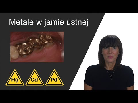
Metale w jamie ustnej
Metale występują w jamie ustnej pod różnymi postaciami. Najczęściej są to wypełnienia amalgamatowe i korony metalowe, lub ceramiczno-metalowe (cienka warstwa ceramiki przykrywającej metal). Spotykane są też w mostach i w protezach. Zalicza się też do nich większość implantów.
Metale te narażone są na ciągłe siły tarcia wiązane z przeżuwaniem, a także na działanie kwasów i na korozję. W wyniku tego przedostają się do śliny, tkanek przyzębia i reszty organizmu.
Wpływ metali na zdrowie
Poza metalami szlachetnymi, takimi jak złoto czy tytan, w stomatologii używa się też metali takich jak srebro, miedź, nikiel. Często występują one jako domieszki.
Metale te organizm zawsze traktuje jako substancje obce i może je tolerować bądź nie, w zależności od wrażliwości układu immunologicznego. W przypadku nietolerancji objawy mogą być różne – począwszy od drobnych stanów zapalnych, które często widoczne są tylko lokalnie w postaci krwawiących dziąseł, aż po poważne alergie lub nawet zaburzenia autoimmunologiczne.
Niestety, przyczyna tych dolegliwości najczęściej nie jest wykrywana i leczenie ogranicza się jedynie do usuwania objawów. Słaba, ale przewlekła aktywacja układu odpornościowego zużywa dużo energii każdego dnia. Może wystąpić wtedy przewlekłe zmęczenie.
Efekty galwaniczne
Ponadto, u pacjentów posiadających kilka źródeł metali w jamie ustnej występują efekty galwaniczne i tzw. efekt anteny.
Efekt galwaniczny (znany również jako ‘efekt baterii’) tworzy się kiedy w jamie ustnej znajdują się dwa lub więcej źródeł różnych metali. Może być to na przykład korona metalowa w pobliżu wypełnienia amalgamatowego. Jony tych metali poruszają się w ślinie tworząc mierzalny prąd. U wrażliwych pacjentów efekt galwaniczny może powodować różne patologiczne zaburzenia, zarówno lokalne jak i ogólne. Przykładem są osłabione reakcje odpornościowe, które mogą być źródłem dyskomfortu odczuwanego w jamie ustnej. Prądy te powodują też przyspieszoną korozję jonów metali. Inne objawy, które zostały powiązane z efektami galwanicznymi to pogorszenie koncentracji i pamięci, bezsenność, bóle i napięcia w klatce piersiowej, palpitacje, szum w uszach i pogorszenie słuchu.
Metale w jamie ustnej działają jako małe ‘anteny’ dla fali elektromagnetycznych wytwarzanych między innymi przez telefony komórkowe i wi-fi. Standardowa absorpcja pól elektromagnetycznych jest zwiększona u pacjentów korzystających z telefonu i jednocześnie mających metale w buzi. Ten efekt może zaburzać też potencjał czynnościowy komórek, powodując nadwrażliwość centralnego układu nerwowego.
Z powodów wyjaśnionych powyżej zrozumiałe jest, że aby zmniejszyć obciążenie układu immunologicznego oraz aby ograniczyć oddziaływanie z polem elektromagnetycznym wszystkie metale powinny być usunięte.
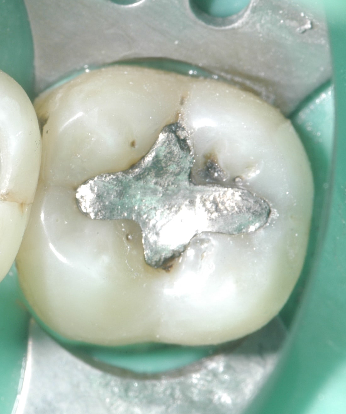
Amalgamat – czym jest?
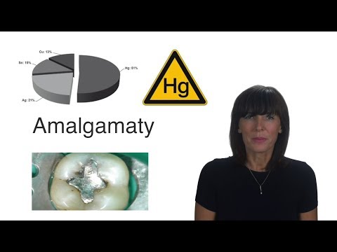
Amalgamat to materiał używany do wypełniania ubytków w zębach. Składa się on w połowie ze stopu metali (najczęściej srebra, miedzi, cyny i cynku) a w połowie z rtęci. Przed użyciem płynna rtęć mieszana jest ze sproszkowanym stopem metali w wyniku czego dochodzi do reakcji innych metali z rtęcią i powstania amalgamatu. Do podstawowych zalet amalgamatu należą łatwość w obsłudze, niska cena i długotrwałość. Należy jednak też zastanowić się nad ich wadami. W 2013 roku weszła w życie międzynarodowa konwencja, mająca na celu ograniczenie użycia rtęci, w tym również amalgamatów, z uwagi na ich trujący wpływ na środowisko po usunięciu. Mając na uwadze ten fakt powinniśmy zastanowić się ponownie nad ich użyciem.
Uwalnianie się rtęci z amalgamatów
Mimo, że związki powstałe w reakcji amalgamacji są dość stabilne, w dalszym ciągu uwalnia się z nich rtęć. Uwalnianie się rtęci wspomagane jest poprzez proces korozji amalgamatów w jamie ustnej i mechaniczne siły tarcia działające na nie (np. mycie zębów, żucie, ścieranie). Szacuje się, że pacjenci z wypełnieniami amalgamatowymi dziennie wchłaniają około 2 mikrogramów rtęci (w zależności od ich ilości). Można zatem nazwać przewlekłym zatruciem o niskiej dawce. Rtęć wchłaniana jest przez błonę śluzową policzków i pod językiem, przedostając się prosto do krwi. Opary rtęci dostają się też do płuc i układu pokarmowego. W kontakcie z bakteriami z jamy ustnej, przewodu pokarmowego oraz krwi, opary przekształcają się w formę organiczną – metylortęć. Metylortęć ma zdolność przenikania przez błony komórkowe i inne bariery takie jak łożysko czy bariera krew- mózg. Dzięki temu ma możliwość przemieszczania się w tkankach.
Toksyczność rtęci
Rtęć jest toksyczna dla wszystkich układów w organizmie. Wdychanie jej oparów negatywnie wpływa m.in na układ nerwowy, pokarmowy, odpornościowy, oddechowy i wydalniczy. Toksyczność rtęci wynika między innymi stąd, że doprowadza ona do zmian w przepuszczalności błon komórkowych, blokuje reakcje enzymatyczne, zaburza przewodzenie impulsów nerwowych, doprowadza do zaburzeń hormonalnych (np. hormonów tarczycy) a nawet może powodować zmiany w kodzie genetycznym.
Głównym celem rtęci jest mózg, rdzeń kręgowy i obwodowy układ nerwowy a zatrucie może prowadzić do zmian neurologicznych i behawioralnych.
Mimo tego, że ilość uwalnianej rtęci z amalgamatów mierzona jest w mikrogramach, nie wolno jej lekceważyć. Wiele badań zaobserwowało wzrost rtęci w krwi i moczu u żywych nośnikach amalgamatów a autopsje wykazały wzrost od 2 do nawet 12-krotny w ilości Hg w różnych tkankach ciała, w tym mózgu.
Podsumowanie
Nie wszyscy doświadczają objawów zatrucia rtęcią, ale każdy kto ma wypełnienia amalgamatowe, ma rtęć w organizmie i narażony jest na codzienne uwalnianie się niej. Efekty mogą być różne, w zależności od ilości amalgamatów, reakcji organizmu i obecności innych źródeł rtęci. Należy pamiętać, że najwięcej rtęci uwalnia się z amalgamatów podczas ich zakładania i usuwania, dlatego niezwykle ważne jest usuwanie ich z odpowiednimi zabezpieczeniami.
Badania i źródła do powyższego tekstu
Amalgamaty
- Bengtsson UG, Hylander LD. Increased mercury emissions from modern dental amalgams. Biometals. 2017;30(2):277–83. doi:10.1007/s10534-017-0004-3
- Mackert JR, Berglund A. Mercury exposure from dental amalgam fillings: absorbed dose and the potential for adverse health effects. Crit Rev Oral Biol Med. 1997;8(4):410– 36.
- Pendergrass JC, Haley BE. Inhibition of brain tubulin-guanosine 5‘-triphosphate interactions by mercury: similarity to observations in Alzheimer‘s diseased brain. Met Ions Biol Syst. 1997;34461–78.
- Eggleston DW, Nylander M. Correlation of dental amalgam with mercury in brain tissue. The Journal of Prosthetic Dentistry. 1987;58(6):704–7. doi:10.1016/0022- 3913(87)90424-0
- Mutter J. Gesund statt chronisch krank!: Der ganzheitliche Weg: Vorbeugung und Heilung sind möglich. 3rd ed. Weil der Stadt: Fit fürs Leben Verlag; 2014. 456 Seiten.
- Taskinen H, Kinnunen E, Riihimäki V. A possible case of mercury-related toxicity resulting from the grinding of old amalgam restorations. Scandinavian Journal of Work, Environment & Health [Internet]. 1989;15(4):302–4. Available from: http://www.jstor.org/stable/40965672
- Bernhoft RA. Mercury toxicity and treatment: a review of the literature. J Environ Public Health. 2012;2012460508. doi:10.1155/2012/460508
- Cariccio VL, Samà A, Bramanti P, Mazzon E. Mercury Involvement in Neuronal Damage and in Neurodegenerative Diseases. Biol Trace Elem Res. 2019;187(2):341–56. doi:10.1007/s12011-018-1380-4
- Ingalls TH. Endemic clustering of multiple sclerosis in time and place, 1934-1984. Confirmation of a hypothesis. Am J Forensic Med Pathol. 1986;7(1):3–8. doi:10.1097/00000433-198603000-00002
- Mutter J. Is dental amalgam safe for humans? The opinion of the scientific committee of the European Commis- sion. Journal of occupational medicine and toxicology (London, England). 2011;62. doi:10.1186/1745-6673-6-2
- Siblerud RL. The relationship between mercury from dental amalgam and the cardiovascular system. Science of The Total Environment. 1990;99(1-2):23–35. doi:10.1016/0048-9697(90)90207-b
- Siblerud RL, Motl J, Kienholz E. Psychometric evidence that mercury from silver dental fillings may be an etiological factor in depression, excessive anger, and anxiety. Psy- chol Rep. 1994;74(1):67–80. doi:10.2466/pr0.1994.74.1.67
- Wojcik DP, Godfrey ME, Christie D, Haley BE. Mercury toxicity presenting as chronic fatigue, memory impairment and depression: diagnosis, treatment, susceptibility, and outcomes in a New Zealand general practice setting (1994- 2006). Neuro Endocrinol Lett. 2006;27(4):415–23.
- Volz U. Qualitative Untersuchungen zur Amalgaminvasion in die Zahnpulpa.: Inaugural-Dissertation zur Erlangung der Doktorwürde. Ulm;1992.
- Christian Nobmann. Die neuen Regelungen zu Amalgam. zm online [Internet]. 2018;(13). Available from: https://www.zm-online.de/archiv/2018/13/titel/die-neuen-regelungen-zu-amalgam/
- Stoiber T, Degen GH, Bolt HM, Unger E. Interaction of mercury(II) with the microtubule cytoskeleton in IMR-32 neuroblastoma cells. Toxicol Lett. 2004;151(1):99–104. doi:10.1016/j.toxlet.2003.11.017
- Stoiber T, Bonacker D, Böhm KJ, Bolt HM, Thier R, Degen GH, Unger E. Disturbed microtubule function and induction of micronuclei by chelate complexes of mercury(II). Mutat Res. 2004;563(2):97–106. doi:10.1016/j.mrgen- tox.2004.06.009
- Pendergrass JC HBE. Mercury-EDTA Complex Specifically Blocks Brain-Tubulin-GTP Interactions: Similarity to Observations in Alzheimer‘s Disease. In Status Quo and Perspective of Amalgam and Other Dental Materials. International Symposium Proceedings. Edited by Friberg LT, Schrauzer GN. Stuttgart: Thieme Verlag;1995 98–105.
- Pendergrass JC, Haley BE. Inhibition of brain tubulinguanosine 5‘-triphosphate interactions by mercury: similarity to observations in Alzheimer‘s diseased brain. Met Ions Biol Syst. 1997;34461–78.
- Barregård L, Svalander C, Schütz A, Westberg G, Sällsten G, Blohmé I, Mölne J, Attman PO, Haglind P. Cadmium,
- mercury, and lead in kidney cortex of the general Swedish population: a study of biopsies from living kidney donors. Environ Health Perspect. 1999;107(11):867–71. doi:10.1289/ ehp.107-1566723
- Drasch G, Schupp I, Riedl G, Günther G. Einfluß von Amalgamfüllungen auf die Quecksilberkonzentration in menschlichen Organen. Dtsch Zahnärztl Z;1992(08):490–6.
- Drasch G, Schupp I, Höfl H, Reinke R, Roider G. Mercury burden of human fetal and infant tissues. European Journal of Pediatrics. 1994;153(8):607–10. doi:10.1007/BF02190671
- Drasch G, Wanghofer E, Roider G. Are blood, urine, hair, and muscle valid bio-monitoring parameters for the internal burden of men with the heavy metals mercury, lead and cadmium? Trace Elem Electrolyt;1997(14):116–23.
- Gottwald B, Traenckner I, Kupfer J, Ganss C, Eis D, Schill WB, Gieler U. „Amalgam disease“–poisoning, allergy, or psychic disorder? Int J Hyg Environ Health. 2001;204(4):223– 9. doi:10.1078/1438-4639-00097
- Guzzi G, Grandi M, Cattaneo C. Should amalgam fillings be removed? Lancet;2002(380):2081.
- Guzzi G, Grandi M, Cattaneo C, Calza S, Minoia C, Ronchi A, Gatti A, Severi G. Dental amalgam and mercury levels in autopsy tissues: food for thought. Am J Forensic Med Pathol. 2006;27(1):42–5. doi:10.1097/01. paf.0000201177.62921.c8
- Levy M, Schwartz S, Dijak M, Weber J-P, Tardif R, Rouah F. Childhood urine mercury excretion: dental amalgam and fish consumption as exposure factors. Environ Res. 2004;94(3):283–90. doi:10.1016/j.envres.2003.07.004
- Lorscheider FL, Vimy MJ, Summers AO. Mercury exposure from „silver“ tooth fillings: emerging evidence questions a traditional dental paradigm. The FASEB Journal. 1995;9(7):504–8. doi:10.1096/fasebj.9.7.7737458
- Kingman A, Albertini T, Brown LJ. Mercury concentrati- ons in urine and whole blood associated with amalgam exposure in a US military population. J Dent Res. 1998;77(3):461–71. doi:10.1177/00220345980770030501
- Mortada W, Sobh M, M El-Defrawy M, E Farahat S. Mercury in dental restoration: Is there a risk of nephrotoxicity? Journal of nephrology. 2002;15171–6.
- Nylander M. MERCURY IN PITUITARY GLANDS OF
- DENTISTS. The Lancet. 1986;327(8478):442. doi:10.1016/ s0140-6736(86)92395-0
- Nylander M, Weiner J. Mercury and selenium concen- trations and their interrelations in organs from dental staff and the general population. Br J Ind Med. 1991;48(11):729– 34. doi:10.1136/oem.48.11.729
- Nylander M, Friberg L, Lind B. Mercury concentrations in the human brain and kidneys in relation to exposure from dental amalgam fillings. Swed Dent J. 1987;11(5):179–87.
- Pizzichini M, Fonzi M, Giannerini F, Mencarelli M, Gasparoni A, Rocchi G, Kaitsas V, Fonzi L. Influence of amalgam fillings on Hg levels and total antioxidant activity in plasma of healthy donors. Science of The Total Environment. 2003;301(1-3):43–50. doi:10.1016/S0048-9697(02)00291-7
- AXELWEINER J, Nylander M. The relationship between mercury concentration in human organs and different predictor variables. Science of The Total Environment.1993;138(1-3):101–15doi:10.1016/0048- 9697(93)90408-X
- Zimmer H, Ludwig H, Bader M, Bailer J, Eickholz P, Staehle HJ, Triebig G. Determination of mercury in blood, urine and saliva for the biological monitoring of an exposure from amalgam fillings in a group with self-reported adverse health effects. Int J Hyg Environ Health. 2002;205(3):205– 11. doi:10.1078/1438-4639-00146
- Takahashi Y. Placental transfer of mercury in pregnant rats which received dental amalgam restorations. Toxicology. 2003;185(1-2):23–33. doi:10.1016/S0300- 483X(02)00588-7
- Ask K, Akesson A, Berglund M, Vahter M. Inorganic mercury and methylmercury in placentas of Swedish women. Environ Health Perspect. 2002;110(5):523–6. doi:10.1289/ehp.02110523
- Holmes AS, Blaxill MF, Haley BE. Reduced levels of mercury in first baby haircuts of autistic children. Int J Toxicol. 2003;22(4):277–85. doi:10.1080/10915810305120
- Morgan DL, Chanda SM, Price HC, Fernando R, Liu J, Brambila E, O‘Connor RW, Beliles RP, Barone S. Disposition of inhaled mercury vapor in pregnant rats: maternal toxicity and effects on developmental outcome. Toxicol Sci. 2002;66(2):261–73. doi:10.1093/toxsci/66.2.261
- Takahashi Y. Release of mercury from dental amalgam fillings in pregnant rats and distribution of mercury in maternal and fetal tissues. Toxicology. 2001;163(2-3):115–26. doi:10.1016/S0300-483X(01)00390-0
- Vahter M, Akesson A, Lind B, Björs U, Schütz A, Berglund M. Longitudinal study of methylmercury and inorganic mercury in blood and urine of pregnant and lactating women, as well as in umbilical cord blood. Environ Res. 2000;84(2):186–94. doi:10.1006/enrs.2000.4098
- Yoshida M, Satoh M, Shimada A, Yamamoto E, Yasutake A, Tohyama C. Maternal-to-fetus transfer of mercury in metallothionein-null pregnant mice after exposure to mercury vapor. Toxicology. 2002;175(1-3):215–22. doi:10.1016/ S0300-483X(02)00084-7
- Yoshida M, Watanabe C, Satoh M, Yasutake A, Sawada M, Ohtsuka Y, Akama Y, Tohyama C. Susceptibility of metall- othionein-null mice to the behavioral alterations caused by exposure to mercury vapor at human-relevant concentration. Toxicol Sci. 2004;80(1):69–73. doi:10.1093/toxsci/ kfh138
- Drasch G, Aigner S, Roider G, Staiger F, Lipowsky G. Mercury in human colostrum and early breast milk. Its dependence on dental amalgam and other factors. J Trace Elem Med Biol. 1998;12(1):23–7.
- Oskarsson A, Schültz A, Skerfving S, Hallén IP, Ohlin B, Lagerkvist BJ. Total and inorganic mercury in breast milk in relation to fish consumption and amalgam in lactating women. Arch Environ Health. 1996;51(3):234–41. doi:10.108 0/00039896.1996.9936021
- Vimy MJ, Hooper DE, King WW, Lorscheider FL. Mercury from maternal “silver” tooth fillings in sheep and human breast milk. Biol Trace Elem Res. 1997;56(2):143–52. doi:10.1007/BF02785388
Tytan
- Safioti LM, Kotsakis GA, Pozhitkov AE, Chung WO, Daubert DM. Increased Levels of Dissolved Titanium Are Associated With Peri-Implantitis – A Cross-Sectional Study. J Periodontol. 2017;88(5):436–42. doi:10.1902/ jop.2016.160524
- Lechner J, Noumbissi S, Baehr V v. Titanium implants and silent inflammation in jawbone-a critical interplay of dissolved titanium particles and cytokines TNF-α and RAN- TES/CCL5 on overall health? EPMA J. 2018;9(3):331–43. doi:10.1007/s13167-018-0138-6
- Delgado-Ruiz R, Romanos G. Potential Causes of Titanium Particle and Ion Release in Implant Dentistry: A Systematic Review. Int J Mol Sci. 2018;19(11). doi:10.3390/ ijms19113585
- Fretwurst T, Nelson K, Tarnow DP, Wang H-L, Giannobile WV. Is Metal Particle Release Associated with Periimplant Bone Destruction? An Emerging Concept. J Dent Res. 2018;97(3):259–65. doi:10.1177/0022034517740560
- Sterner T, Schütze N, Saxler G, Jakob F, Rader CP. Auswirkungen von klinisch relevanten Aluminium Keramik-, Zirkonium Keramik- und Titanpartikel unterschiedlicher Grösse und Konzentration auf die TNFalpha-Ausschüttung in einem humanen Makrophagensystem [Effects of clinically relevant alumina ceramic, zirconia ceramic and titanium particles of different sizes and concentrations on TNF-alpha release in a human macrophage cell line]. Biomed Tech (Berl). 2004;49(12):340–4. ger. doi:10.1515/ BMT.2004.063
- Rader CP, Sterner T, Jakob F, Schütze N, Eulert J. Cytokine response of human macrophage-like cells after contact with polyethylene and pure titanium particles. The Journal of Arthroplasty. 1999;14(7):840–8. doi:10.1016/ S0883-5403(99)90035-9
- Cadosch D, Chan E, Gautschi OP, Meagher J, Zellweger R, Filgueira L. Titanium IV ions induced human osteoclast differentiation and enhanced bone resorption in vitro. J Biomed Mater Res A. 2009;91(1):29–36. doi:10.1002/ jbm.a.32183
- Volker von Baehr, Sabine Schütt. lmmunologische Grundlagen der Titan-induzierten Periimplantitis. ZMK. 2011;2721–6.
- Baehr V v. Titanunverträglichkeit. ZWR. 2018;127(04):180– 1. doi:10.1055/a-0563-2511
- Schütt S, Von Baehr V. Hyperreaktivität von Gewebemakrophagen nach Kontakt mit Titanoxidpartikeln als Ursache einer verstärkten lokalen Entzündungsreaktion bei Patienten mit Periimplantitis. ZWR – Das Deutsche Zahnärzteblatt;2010(119):222–32.
- Lindhe J, Meyle J. Peri-implant diseases: Consensus Report of the Sixth European Workshop on Periodontology. J Clin Periodontol. 2008;35(8 Suppl):282–5. doi:10.1111/ j.1600-051X.2008.01283.x
- Barão VAR, Yoon CJ, Mathew MT, Yuan JC-C, Wu CD, Sukotjo C. Attachment of Porphyromonas gingivalis to corroded commercially pure titanium and titanium-aluminumvanadium alloy. J Periodontol. 2014;85(9):1275–82. doi:10.1902/jop.2014.130595
- Apaza-Bedoya K, Tarce M, Benfatti CAM, Henriques B, Mathew MT, Teughels W, Souza JCM. Synergistic interactions between corrosion and wear at titanium-based dental implant connections: A scoping review. J Periodont Res. 2017;52(6):946–54. doi:10.1111/jre.12469
- Senna P, Antoninha Del Bel Cury A, Kates S, Meirelles L. Surface Damage on Dental Implants with Release of Loose Particles after Insertion into Bone. Clin Implant Dent Relat Res. 2015;17(4):681–92. doi:10.1111/cid.12167
- Olmedo D, Fernández MM, Guglielmotti MB, Cabrini RL. Macrophages related to dental implant failure. Implant Dent. 2003;12(1):75–80. doi:10.1097/01. id.0000041425.36813.a9
- Hallab NJ, Jacobs JJ. Biologic effects of implant debris. Bull NYU Hosp Jt Dis. 2009;67(2):182–8.
- Jacobi-Gresser E. Pathogenese der Periimplantitis. Dentale Implantologie & Parodontologie [Internet];08.2019. Available from: https://www.dimagazin-aktuell.de/implantologie/periimplantitis/story/pathogenese-der-periimplantitis__6705.html
- Hedenborg M. Titanium dioxide induced chemiluminescence of human polymorphonuclear leukocytes. International Archives of Occupational and Environmental Health. 1988;61(1):1–6. doi:10.1007/BF00381600
- Stejskal VDM, Danersund A, Lindvall A, Hudecek R, Nordman V, Yaqob A, Mayer W, Bieger W, Lindh U. Metal-specific lymphocytes: biomarkers of sensitivity in man. Neuro Endocrinol Lett. 1999;20(5):289–98.
- McGuff HS, Heim-Hall J, Holsinger FC, Jones AA, O‘Dell DS, Hafemeister AC. Maxillary osteosarcoma associated with a dental implant: report of a case and review of the literature regarding implant-related sarcomas. J Am Dent Assoc. 2008;139(8):1052–9. doi:10.14219/jada. archive.2008.0307
- Poggio CE. Plasmacytoma of the mandible associated with a dental implant failure: a clinical report. Clin Oral Implants Res. 2007;18(4):540–3. doi:10.1111/j.1600-0501.2007.01361.x
- Dib LL, Soares AL, Sandoval RL, Nannmark U. Breast metastasis around dental implants: a case report. Clin Implant Dent Relat Res. 2007;9(2):112–5. doi:10.1111/j.1708-8208.2007.00033.x
- Weingart D, Steinemann S, Schilli W, Strub JR, Hellerich U, Assenmacher J, Simpson J. Titanium deposition in regional lymph nodes after insertion of titanium screw implants in maxillofacial region. International Journal of Oral and Maxillofacial Surgery. 1994;23(6):450–2. doi:10.1016/S0901- 5027(05)80045-1
- Fujii Y. Sensation of Balance Dysregulation Caused/
- Aggravated by a Collection of Electromagnetic Waves in a Dental Implant. OJAPr. 2014;02(03):29–35. doi:10.4236/ ojapr.2014.23004
Inne metale
- Saravanakumar P, Thallam Veeravalli P, Kumar V A, Mohamed K, Mani U, Grover M, Thirumalai Thangarajan S. Effect of Different Crown Materials on the InterLeukin-One Beta Content of Gingival Crevicular Fluid in Endodontically Treated Molars: An Original Research. Cureus. 2017;9(6):e1361. doi:10.7759/cureus.1361
- Stejskal J, Stejskal V. The role of metals in autoimmunity and the link to neuroendocrinology. Neuro endocrinology letters. 1999;20351–64.
- Lehmann I, Sack U, Lehmann J. Metal ions affecting the immune system. Met Ions Life Sci. 2011;8157–85.
- Mutter J, Klinghardt D. Amalgam: Risiko für die Menschheit; Quecksilbervergiftungen richtig ausleiten, neue Fakten und Hilfe, auch nach der Amalgamentfernung! 3rd ed. Weil der Stadt: Fit-fürs-Leben-Verl. in der NaturaViva-Verl.-GmbH; 2013. 169 p. (Gesundheit).
- Dr. Kurt E. Müller. Immunreaktion auf physiologisch
- nicht benötigte Metalle. UMG [Internet];2013(4). Available from: https://www.deguz.de/de/fachinformationen/metalle-und-metallischer-zahnersatz/immunreaktion-auf-physiologisch-nicht-benoetigte-metalle/
- Khan M, Naqvi AH, Ahmad M. Comparative study of the cytotoxic and genotoxic potentials of zinc oxide and titanium dioxide nanoparticles. Toxicol Rep. 2015;2765–74. doi:10.1016/j.toxrep.2015.02.004
- Bjorklund G, Stejskal V, Urbina MA, Dadar M, Chirumbolo S, Mutter J. Metals and Parkinson‘s Disease: Mechanisms and Biochemical Processes. Curr Med Chem. 2018;25(19):2198–214. doi:10.2174/092986732566617112912 4616
- Thier R, Bonacker D, Stoiber T, Böhm KJ, Wang M, Unger E, Bolt HM, Degen G. Interaction of metal salts with cytoskeletal motor protein systems. Toxicol Lett. 2003;140- 14175–81.doi:10.1016/S0378-4274(02)00502-7
- Becker K, Kaus S, Krause C, Lepom P, Schulz C, Seiwert M, Seifert B. German Environmental Survey 1998 (GerES III): environmental pollutants in blood of the German population. Int J Hyg Environ Health. 2002;205(4):297–308. doi:10.1078/1438-4639-00155
- Becker K, Schulz C, Kaus S, Seiwert M, Seifert B. German Environmental Survey 1998 (GerES III): environmental pollutants in the urine of the German population. Int J Hyg Environ Health. 2003;206(1):15–24. doi:10.1078/1438-4639- 00188
- Zohdi H, Emami M, Reza H. Galvanic Corrosion Behavior of Dental Alloys. In: Valdez B, editor. Environmental and Industrial Corrosion – Practical and Theoretical Aspects: InTech; 2012.
- Procházková J, Podzimek S, Tomka M, Kucerová H, Mihaljevic M, Hána K, Miksovský M, Sterzl I, Vinsová J. Metal alloys in the oral cavity as a cause of oral discomfort in sensitive patients. Neuro Endocrinol Lett. 2006;27 Suppl 153– 8.
- Johansson BI. Electrochemical action due to short-circuiting of dental alloys. An in vitro and in vivo study. Swed Dent J Suppl. 1986;331–47.
- Ciszewski A, Baraniak M, Urbanek-Brychczyńska M. Corrosion by galvanic coupling between amalgam and different chromium-based alloys. Dent Mater. 2007;23(10):1256–61. doi:10.1016/j.dental.2006.11.006
- Taher NM, Al Jabab AS. Galvanic corrosion behavior of implant suprastructure dental alloys. Dent Mater. 2003;19(1):54–9.
- Dr. med. dent. Johann Lechner. Immunstress durch Zahnmetalle und Elektrosmog. Raum&Zeit;1995(74):5–13.
- Virtanen H, Huttunen J, Toropainen A, Lappalainen R. Interaction of mobile phones with superficial passive metallic implants. Phys Med Biol. 2005;50(11):2689–700. doi:10.1088/0031-9155/50/11/017